Optical vortex beams are a type of beam that possess quite a unique wavefront. Most beams that are analysed in the laboratory will exhibit a wavefront that is either flat or radially symmetric. Highly aberrated beams will show more complex wavefronts but an optical vortex beam will exhibit a distinct wavefront in which the phase is constant along the angular coordinate. Such a phase pattern resembles a fluid vortex, or whirlpool, as they occur in sinking fluids and hence the name.
To impart such a distinct phase pattern into the beam’s wavefront, a filter-like optical element is needed. Such an element is referred to as an optical vortex phase plate. The surface of this element could be described as something akin to a spiral staircase in which each step imparts a different optical path difference. For this reason, the optical vortex phase plate is also known as a spiral phase plate.
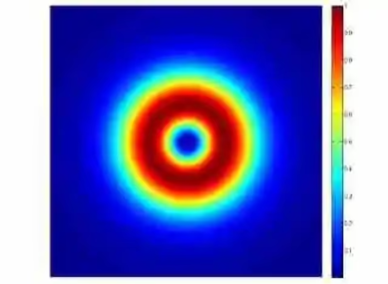
When the optical vortex beam is brought to a focus with a positive lens, the focused spot is not the common Gaussian beam that is expected from conventional laser beams. Instead, it is characterized by a ring structure surrounding a region of zero irradiance. Some defined combination of Laguerre-Gaussian modes could also give rise to such an irradiance pattern, but it is much more practical to use an optical vortex phase plate when this sort of beam pattern is needed. Optical vortex beams are more than a mere curiosity as they can find applications in many fields. In the newly emerging field of quantum computation, for instance, a beam with a phase vortex is said to possess an angular momentum, also referred to as a topological charge, which can be then used to encode quantum information. In fluorescence microscopy, an optical vortex phase beam can be used to help the imaging system achieve a resolution beyond the diffraction limit specified for the operating wavelength. In a nutshell, this is accomplished by illuminating the sample with two different beams of different wavelengths. One of the beams is from a wavelength that will induce the desired fluorescence. The other beam is from a different wavelength and an optical vortex phase plate is placed along its path. When the two beams coincide in the sample, the ring structure from the optical vortex phase beam encloses an area that turns out to be smaller than the area defined by the diffraction limit of the fluorescence wavelength. This technique is known as stimulated emission depletion (STED) microscopy.